
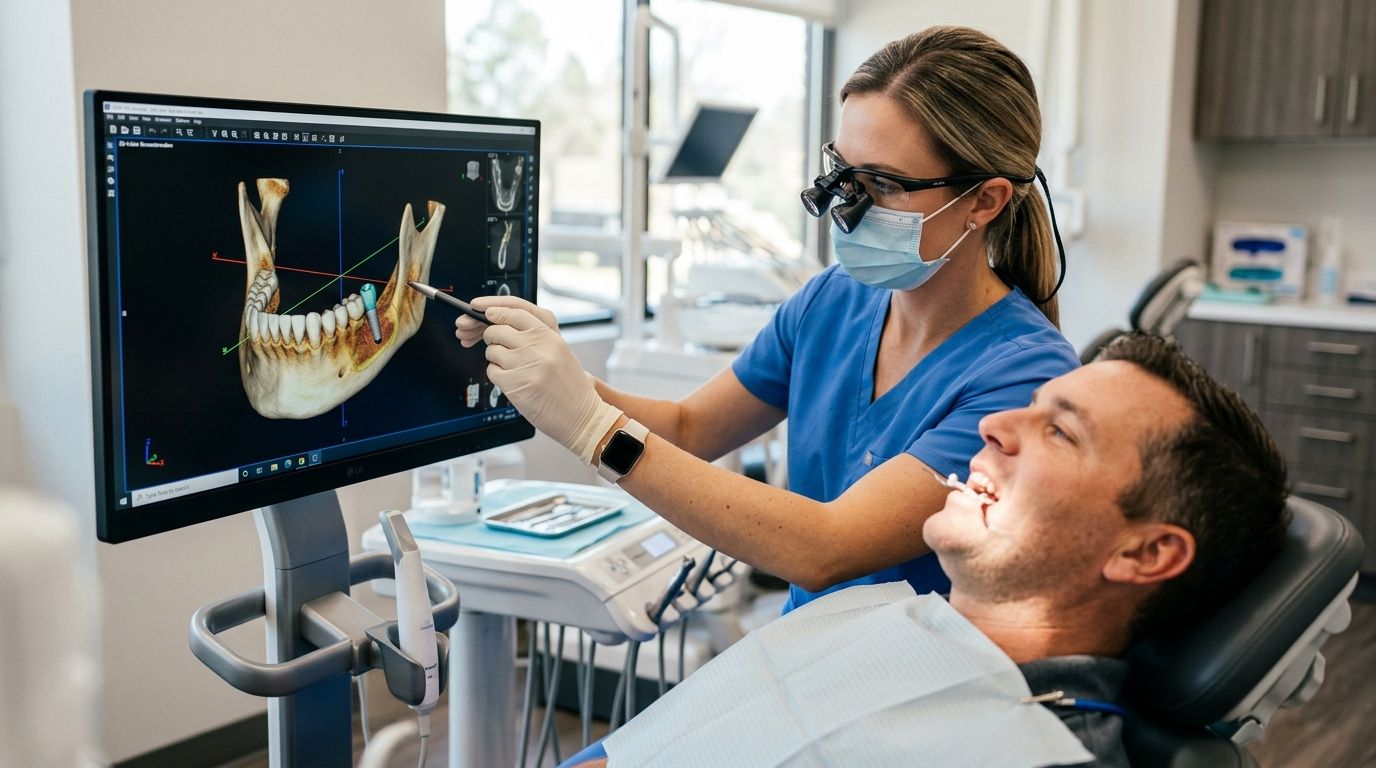
3D imaging plans a dental implant by pairing a cone beam CT (CBCT) scan with digital planning software, so your dentist can see bone density, nerve position, and sinus location before touching a drill. The plan is then built virtually — implant depth, angle, and size are set on screen and often transferred to your mouth through a printed surgical guide. This is the planning method Dental Arts of Atlantis uses for implant cases in 2026, from a single missing tooth to a full-arch case. What the headline description leaves out: the scan often uncovers bone loss that adds a grafting stage before any implant goes in.
- 3D imaging dental implant planning combines a CBCT scan with digital software to map bone, nerves, and sinus location before surgery.
- Dental Arts of Atlantis uses this planning method in 2026 for single-tooth implants through full-arch cases.
- A surgical guide built from the 3D plan transfers the on-screen implant angle and depth into your mouth.
- Patients with low bone density get flagged for grafting before the implant stage, not mid-surgery.
- The scan takes a few minutes chairside and is ordered even for a single, simple-looking implant site.
Why this matters
A standard 2D dental X-ray shows height and width. It cannot show depth, which means it cannot reliably show how close an implant site sits to the inferior alveolar nerve or the sinus floor. Placing an implant off a flat image means planning blind on the one dimension that matters most for safety.
CBCT imaging fixes that by capturing a full 3D volume of your jaw in one pass. Your dentist can rotate the image, measure bone in millimeters at the exact planned site, and catch problems — thin bone, a low sinus, a nerve running closer to the ridge than expected — before surgery day instead of during it. That is the difference between a first dental implant consultation that ends with a real surgical plan and one that ends with a guess.
In 2026, a guided implant plan is the standard of care for any case where the nerve canal or sinus floor sits near the planned site.
How to use 3D imaging to plan a dental implant
The workflow runs in six steps, and each one feeds the next.
- CBCT scan. You sit or stand in the imaging unit for a few minutes while it captures a 360-degree volume of your jaw, teeth, and surrounding structures.
- Digital surface scan. A handheld intraoral scanner — or a traditional impression — captures the exact shape of your teeth and gums.
- Software merges the two data sets. Planning software overlays the surface scan onto the CBCT volume, producing one model that shows bone underneath and tooth position above.
- Virtual implant placement. Your dentist positions a to-scale digital implant at the site, checking angle, depth, and clearance from the nerve canal and sinus floor.
- Surgical guide design. Once the plan is approved, a guide is designed to seat over your teeth or gums and hold the drill at the exact angle and depth set on screen.
- Guided placement. On surgery day, the guide transfers the digital plan into your mouth instead of relying on freehand judgment alone.
The steps are identical whether you are replacing one missing tooth or rebuilding a full arch. What changes is how much bone gets evaluated and how many sites have to be checked against each other.
2D X-ray vs. CBCT for implant planning
| Factor | 2D dental X-ray | CBCT 3D scan |
|---|---|---|
| Dimensions captured | Height and width | Height, width, and depth |
| Nerve canal position | Approximate, can be obscured | Measurable at the planned site |
| Sinus floor clearance | Hard to judge | Measurable in millimeters |
| Bone density reading | Limited | Site-specific |
| Drives a surgical guide | No | Yes |
| Best for | Routine checkups and cavity detection | Implant planning, extractions near nerves |
Honest limits of CBCT: it costs more than a routine X-ray, it delivers more radiation than a single bitewing film, and it adds a planning step before surgery. For a routine cleaning it is unnecessary. For an implant, that extra step is what keeps a drill away from a nerve.
Single-tooth implant: one site, full 3D check
A single implant still needs the whole workflow. The nerve canal and sinus floor sit in different positions from one patient to the next, so "simple case" is a conclusion the scan produces, not an assumption you start with. The plan sets one implant angle, one depth, and confirms there is enough bone width on both sides of the planned fixture.
Verdict: get the 3D scan even for one tooth.
Full-arch implants: multiple sites checked against each other
Full-arch planning adds a layer. Each implant position has to work with the others and with the final bite, not just with the bone at its own site. That is why All-on-4 implants planning leans so heavily on the digital model — angled implants placed to reach denser bone only work if the angles are set before surgery, not improvised during it.
Verdict: 3D planning is non-negotiable for a full arch.
Start your implant plan with a 3D scan
See your bone, nerve, and sinus position mapped before surgery day.
Why the 3D implant plan varies from patient to patient
No two CBCT scans read the same way. The plan that comes out of one shifts based on a short list of factors:
- Bone density and volume at the site. Thin or soft bone changes implant size, angle, or whether grafting comes first.
- Proximity to the nerve or sinus. A lower-jaw implant near the nerve canal, or an upper-jaw implant near the sinus floor, needs tighter depth control.
- Number of implants needed. One site is a single check; a full arch means every position has to agree with every other.
- Existing bone loss. Years of a missing tooth or untreated gum disease resorb the ridge, and that shows clearly on the 3D volume.
- Bone-affecting medical conditions. Osteoporosis changes how bone heals around an implant and factors into the timeline the plan builds in.
- Smoking history. Smoking slows healing at the implant site, which makes the plan more conservative on load timing.
Bone that looks adequate on a flat X-ray sometimes reads as too thin once the 3D volume is measured. That is why bone grafting for low bone density gets written into the plan up front for those patients instead of discovered halfway through surgery.
“A surgical guide is only as accurate as the scan it was built from.”
Does 3D imaging replace the surgical plan discussion?
No. 3D imaging dental implant planning produces the data; the treatment plan is still a conversation about timeline, healing, staging, and what happens if the scan shows grafting is needed first. Dental Arts of Atlantis builds the imaging into a comprehensive plan rather than handing you a scan and a quote.
What if the scan shows there is not enough bone?
If the scan shows there is not enough bone, the plan adds a grafting or sinus-lift stage before implant placement — the implant is not cancelled, it is sequenced. Finding this on the screen in 2026 rather than in the chair is the entire point of scanning first.
How soon before surgery should the scan happen?
The scan happens at or near the consultation stage, because the surgical guide cannot be designed until the digital implant position is approved. Anatomy can also change after an extraction as the site heals, so timing gets set case by case.
FAQ
What is 3D imaging dental implant planning?
It is the use of a CBCT scan plus digital planning software to set an implant’s position, angle, and depth before surgery. The approved plan is often transferred to your mouth through a surgical guide.
What’s the difference between a CBCT scan and a regular dental X-ray?
A regular dental X-ray is a flat 2D image showing height and width only. A CBCT scan captures a full 3D volume, adding depth so bone, nerve position, and sinus clearance can be measured at the planned implant site.
How much does 3D imaging for implant planning cost?
Pricing depends on the case and on how the imaging is billed, so ask the office for a current estimate before booking. Whether your plan covers diagnostic imaging tied to an implant also varies by insurer.
Can 3D imaging show if I need a bone graft before implants?
Yes. A CBCT scan measures bone density and volume at the exact planned site, which is how low bone density gets caught before surgery instead of during it. That finding is what routes a patient to grafting first.
Is 3D imaging safe if I’m pregnant?
Elective dental imaging is generally deferred during pregnancy unless there is an urgent dental problem. Tell your dentist you’re expecting so timing and alternatives get reviewed first.
Do I need 3D imaging for a single implant or only for full-arch cases?
Both. A single-tooth case still needs nerve and sinus clearance confirmed at that one site, while a full-arch case adds the step of checking how multiple implant positions relate to each other.
How is a surgical guide made from the scan?
After the digital implant position is approved in the software, a guide is designed to seat over your teeth or gums and hold the drill at the planned angle and depth. It is fabricated before surgery day, not during it.
Is 3D implant planning worth it for a straightforward case?
Yes, because "straightforward" is a conclusion the scan reaches, not a starting assumption. Nerve canals and sinus floors sit in different positions in every patient regardless of how simple the case looks.
One last thing
The most useful thing a CBCT scan does is not always the implant plan itself — it is the incidental finding. A scan ordered to measure one implant site also shows retained root fragments, unerupted teeth, and sinus changes that never appeared on a 2D film. If you are weighing an implant against saving the tooth you still have, get the 3D scan first: it sometimes shows enough remaining root structure to make saving the tooth the better call in 2026, which is the one outcome no implant plan can give you back later.
Related guides
- How to prepare for your first dental implant consultation
- All-on-4 implants for denture wearers switching to fixed teeth
- Bone grafting for dental implants with low bone density
- Dental implants for one missing tooth
- How to save a tooth instead of getting it extracted